



Can AI-powered eye scans detect Alzheimer’s and Parkinson’s early?
AI-assisted OCT may reveal retinal changes linked to Alzheimer’s and Parkinson’s, but experts say eye scans cannot yet diagnose either condition alone.


By Ahmet Taş | Wise News Press
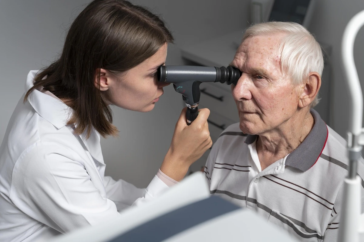
IZMIR, Türkiye — AI-assisted optical coherence tomography may reveal retinal changes associated with Alzheimer’s and Parkinson’s disease, but current evidence does not support using a rapid eye scan as a stand-alone diagnostic test.
Researchers are studying whether subtle changes in retinal nerve layers and small blood vessels could provide early clues about neurodegenerative diseases before symptoms such as memory loss, tremor or impaired balance become prominent. Specialists caution, however, that similar retinal findings can occur with aging, diabetes, glaucoma, high blood pressure and other medical conditions.
Why the retina may reflect brain health
The retina is the light-sensitive neural tissue at the back of the eye. It receives visual information and transmits signals to the brain through the optic nerve. Because the retina is closely connected to the central nervous system, researchers have increasingly examined whether changes in the brain may also leave measurable signs in the eye.
Studies of Alzheimer’s disease have investigated thinning of the retinal nerve fiber layer, changes in ganglion cells and differences in the retina’s microvascular network. Parkinson’s research has examined similar structural and vascular features, although findings have not been consistent across all patient groups.


A 2026 systematic review of OCT research in preclinical and early Alzheimer’s disease found that retinal measurements could become useful complementary biomarkers. The authors nevertheless reported substantial variation between studies and concluded that imaging methods and patient-selection criteria must be standardized before retinal screening can be widely adopted in clinical practice.
How an OCT eye scan works
Optical coherence tomography, commonly known as OCT, uses reflected light to create high-resolution, cross-sectional images of the retina. The technology allows clinicians to examine the retinal layers, macula, optic nerve head and retinal nerve fiber layer at a microscopic scale.
The examination is generally non-invasive, does not involve ionizing radiation and normally requires no physical contact with the eye. The patient looks into the device while a series of images is captured within seconds.


OCT is already widely used in ophthalmology to assess conditions including glaucoma, age-related macular degeneration, diabetic retinopathy and diseases affecting the optic nerve. It provides structural information, while OCT angiography can also visualize blood flow within the retina’s small vessels without injecting contrast dye.
The speed of the scan has led to claims that dementia could eventually be identified through a “one-minute eye test.” The image-acquisition process may be brief, but interpreting the findings and determining their medical significance is considerably more complex.
What artificial intelligence looks for
Artificial intelligence systems can examine thousands of measurements across retinal images. Algorithms may analyze the thickness and symmetry of retinal layers, the shape of the optic nerve, vessel density, branching patterns and features that may be difficult to identify through routine visual inspection.
Researchers train these systems using images from people with diagnosed neurological diseases and control groups. The resulting model is then tested to determine whether it can distinguish between the groups in previously unseen images.
A 2025 systematic review and meta-analysis evaluated 10 studies using AI-assisted retinal imaging for Alzheimer’s, Parkinson’s and related neurodegenerative conditions. The pooled diagnostic performance produced an area under the curve of 0.73, indicating moderate rather than definitive accuracy. Performance was measured at 0.72 for Alzheimer’s and 0.70 for Parkinson’s, while considerable variation was found among the included studies.
The review’s authors said the results demonstrated promise but called for large prospective studies, standardized imaging protocols and more consistent patient-selection methods. Models developed using one hospital, camera type or patient population may not perform equally well in another clinical setting.
Can an eye scan diagnose Alzheimer’s disease?
An OCT scan cannot currently confirm that a person has Alzheimer’s disease. Retinal findings are not sufficiently specific, and a normal scan cannot reliably rule the disease out.
Assoc. Prof. Uğur Ünsal, an ophthalmology specialist at Batıgöz Health Group in İzmir, said AI-supported retinal analysis should be viewed as a potentially useful risk-assessment tool rather than a definitive diagnostic method. Ünsal specializes in eye diseases and vitreoretinal surgery at the group’s Çankaya branch.
“These systems should be regarded as supportive technologies that may contribute to risk assessment.”
When suspicious findings are detected, patients still require a broader medical evaluation. According to the U.S. National Institute on Aging, doctors assess Alzheimer’s disease through medical history, cognitive and language testing, evaluation of daily functioning, laboratory investigations and, when appropriate, brain imaging or measurements of disease-related proteins in blood or cerebrospinal fluid.
No single biomarker independently provides a complete dementia diagnosis. Biomarkers are interpreted alongside symptoms, clinical examination and other test results.
Can retinal imaging identify Parkinson’s disease?
The same limitations apply to Parkinson’s disease. Researchers have found possible associations between Parkinson’s and changes in retinal nerve layers, visual processing and retinal blood vessels, but OCT is not an approved stand-alone diagnostic test for the condition.
Parkinson’s remains primarily a clinical diagnosis based on a person’s medical history, symptoms and neurological examination. Doctors particularly assess slowness of movement, resting tremor, muscle rigidity and balance problems.
Tests such as MRI, dopamine transporter imaging, blood analysis or skin biopsy may support an evaluation or help exclude conditions that resemble Parkinson’s. The Parkinson’s Foundation states that no single laboratory or imaging test currently confirms Parkinson’s disease on its own.
This is important because an AI model may recognize patterns associated with a group of Parkinson’s patients without proving that those patterns are unique to Parkinson’s or capable of predicting the disease in healthy individuals.
Why early detection remains a major goal
Alzheimer’s and Parkinson’s can begin affecting the nervous system years before their most recognizable symptoms appear. Researchers therefore aim to identify reliable biological changes during earlier stages, when monitoring, treatment planning and participation in clinical studies may be more useful.
A safe and accessible retinal test would offer practical advantages. OCT machines are already present in many eye clinics, the scan is fast and repeat examinations can be performed without exposing patients to radiation.
Retinal imaging may eventually help identify people who should receive additional cognitive testing, neurological examination or established biomarker assessments. It could also contribute to monitoring disease progression or evaluating responses to treatment.
The current evidence does not support offering OCT to symptom-free people as a definitive Alzheimer’s or Parkinson’s screening test. The technology remains an active area of research, and commercial claims promising an immediate dementia diagnosis from a brief eye scan should be treated cautiously.
People experiencing worsening memory problems, repeated confusion, difficulty carrying out familiar tasks, changes in personality, unexplained tremor, muscle stiffness, slowed movement or balance difficulties should seek an appropriate medical assessment rather than relying on a retinal scan alone.
OCT may become one component of a broader early-detection system, particularly when combined with cognitive testing, blood biomarkers and neurological evaluation. For now, its role in Alzheimer’s and Parkinson’s care remains supportive and investigational rather than diagnostic.
WiseNewsPress.com
What's Your Reaction?
 Like
0
Like
0
 Dislike
0
Dislike
0
 Love
0
Love
0
 Funny
0
Funny
0
 Wow
0
Wow
0
 Sad
0
Sad
0
 Angry
0
Angry
0

Editor | Wise News Press — Delivering accurate, timely global news with integrity, insight, and editorial responsibility.





















Comments (0)